
Case Study: Treating Prostate Cancer with NanoKnife Focal Therapy
Patient Background
A male patient in his 60s was first diagnosed with low-risk prostate cancer (Gleason 6) more than 10 years ago. Because this type of cancer often grows slowly, he was managed with active surveillance, a strategy that involves regular PSA tests, MRI scans and biopsies to monitor the cancer without immediate treatment.
For a decade, his cancer remained stable.
However, in 2024, his prostate MRI found a new lesion. Targeted prostate biopsy showed the cancer had progressed to Gleason 7 disease, indicating a more aggressive tumour that required treatment.
Choosing a Treatment Approach
At this stage, several treatment options were discussed, including:
Continuing active surveillance (not recommended)
Radical prostatectomy (surgical removal of the prostate)
Radiation therapy
Focal therapy using NanoKnife
Because the cancer was localised to a specific region of the prostate, focal therapy was considered an appropriate option. This approach treats the tumour while preserving the surrounding healthy prostate tissue, helping reduce the risk of urinary and sexual side effects.
The patient elected to proceed with NanoKnife focal therapy.
The Procedure
In 2025, the patient underwent NanoKnife (irreversible electroporation) focal therapy to treat the tumour located in the left posterior part of the prostate.
NanoKnife uses short electrical pulses to destroy cancer cells while preserving nearby structures such as nerves and blood vessels. This precision allows effective cancer treatment while minimising damage to surrounding tissue.
The procedure was completed successfully. The patient went home on the same day with an urinary catheter. The catheter was removed 5 days later without issues.
MRI Pre and Post NanoKnife Treatment
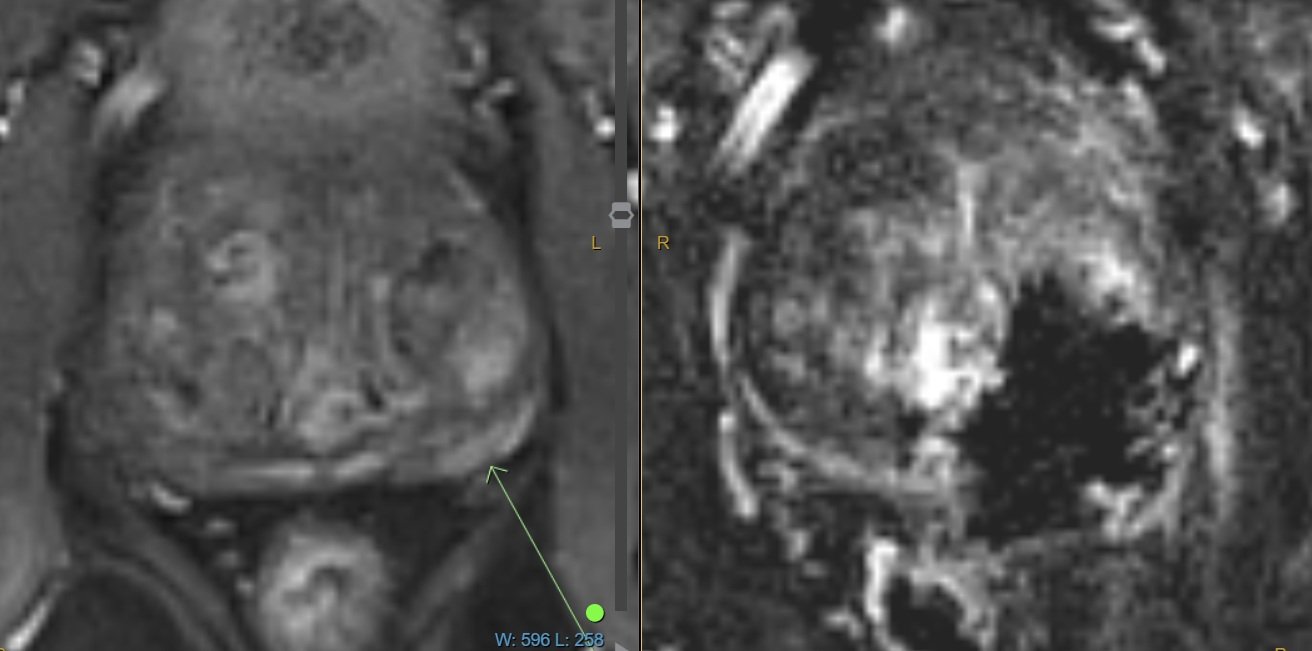
Left: MRI pre-treatment showing the prostate tumour.
Right: MRI 2 weeks post-treatment showing the treatment coverage is satisfactory.
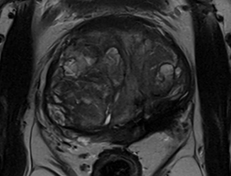
MRI 6 months post-treatment. The cancerous tissue is replaced with fibrosis (scar tissue), with no new pathology identified.
Follow-Up Results
PSA reduced by 50%
MRI in January 2026 showed resolution of the treated tumour with no new suspicious lesions
Repeat prostate biopsy in February 2026 showed fibrosis and degenerative treatment changes in the treated area, with no malignancy identified.
No incontinence
Nomal erectile function
Mild reduction of semen volume
Ongoing Monitoring
Even after successful focal therapy, ongoing monitoring remains important.
The follow-up plan includes:
6 monthly PSA testing
Prostate MRI every 1-2 years, or sooner if there is any clinical concerns
± Biopsy if MRI shows abnormal changes in the future
Key Takeaways
This case highlights several important points:
Low risk prostate cancers can be safely monitored for years with active surveillance
If the cancer progresses, focal therapy can offer an effective treatment option for selected patients
NanoKnife targets the tumour while minimising impact on continence and erectile function
Careful follow-up with PSA testing and MRI remains important
*This case is presented for educational purposes. Individual results vary and treatment decisions should be made in consultation with a qualified specialist.